


| Radiology Room |
| Ultrasound Room |
| Surgery Room |
| Laboratory Room |
| Comprehensive Room |
| Pediatrics Room |
| Dental Room |
| Medical operation instruments |
| Hospital Furniture |
| Medical supplies |


News Center
3d Ultrasound: Capturing the Precious Moments of Pregnancy
Nowadays, many hospitals and clinics provide 3d ultrasound services to pregnant women. Pregnant women can also obtain ultrasound services from 3d ultrasound centers. Maternity hospitals, clinics and other medical facilities are embracing ultrasound technology due to the growing need among pregnant women to know the health state of their developing babies and the sex of their babies.
A mother is able know the health of the developing fetus by use of a 3d ultrasound device. With this device, doctors can be able to discover at an early stage if the developing baby has any health complications. Use of ultrasounds to check the health condition of a baby is called fetal anomaly scanning. This scanning will reveal if a baby has heart anomalies, brain anomalies among other anomalies. In the near future, elective ultrasound devices will be able to detect whether a baby has cerebral palsy.
3d ultrasounds are also normally used by many pregnant women to determine the sex of their babies. The sex of a baby can be determined with 95% accuracy using an ultrasound machine during the 20th week of the pregnancy. Determining the sex of a baby using an ultrasound machine is best done between the 20th and the 32nd week of the pregnancy. Above the 32nd week, there is a possibility that the baby may have descended down to the pelvis and it will be hard to determine the baby’s gender using an ultrasound device.
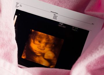
Three Dimensional Ultrasound Photo
Medical literature however reports that it is possible to determine the sex of the baby using an ultrasound as early as the 15th week of the pregnancy. The success rate at the 15th week of the pregnancy is 50%. One will have to pay for the ultrasound scanning whether it succeeds or not. In case the scanning fails to determine the sex of the baby during the 15th week, the mother will have to wait until the 17th week and carry out another scanning. From the 17th week to the 32nd week of the pregnancy, it is possible to determine the gender of a baby with an accuracy level exceeding 90%.
Apart from being used to determine the sex and health state of a baby, 3d ultrasound scanning can simply be used to see how the baby looks like and capture the baby’s moments in his/her mother’s womb. The best images of a baby’s looks can be captured between the 28th and the 32nd week of the pregnancy. Once the mother has been able to see how her baby looks like, she will be utterly convinced that she is carrying a real human being in her womb.
Most providers of 3d ultrasound services will offer moms-to-be services that will enable them capture the images of their babies. These images can be stored for memory. The images of the developing baby in the womb are precious images that should be preserved. Ultrasound centers normally use high quality equipments that will enable them to capture the best images of the developing fetus. A well experienced ultrasonographer/ultrasound technician will provide high quality images.
New advancements in the 3d ultrasound niche, has made it possible to watch a baby’s moves real time via a large screen. This can later be recorded and stored in a DVD for later viewing. With this advancement, it is now possible to show pregnant women contemplating divorce the real time movements of their babies’ in their wombs. After seeing these, these mothers will be convinced that what they are carrying in their wombs are real baby’s who have real lives. These may influence them to rethink their decision to carry out abortion. 3d ultrasound has been used by many pro life organizations to convince pregnant women not to carry out abortion. Many charitable organizations in America pay the fees for ultrasound scanning for women contemplating divorce.
The pregnancy period is a precious time not only to the pregnant mother-to-be but also to the baby in the womb. Capturing images of the developing baby using 3d ultrasound will enable the precious moments of the developing fetus to be observed and stored. These moments can be stored in 3d pictures and DVD’s.
